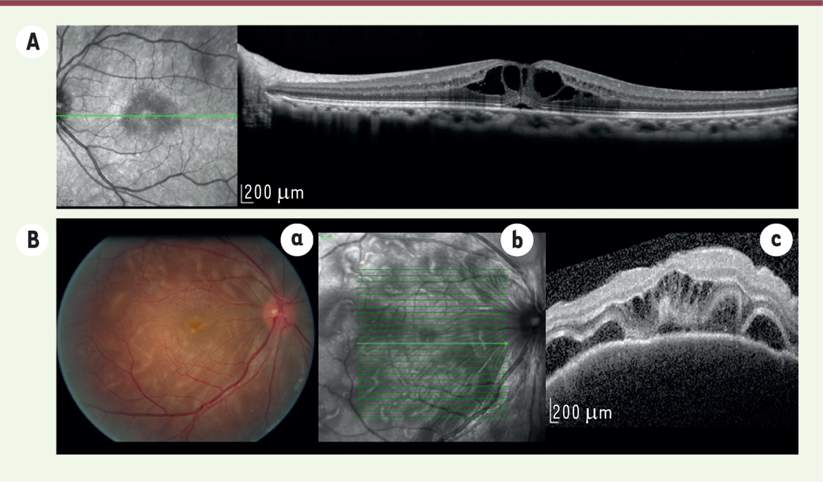
Figure 1.

Télécharger l'image originale
A. Image infrarouge et OCT (optical coherence tomography, Spectralis, Heidelberg, Allemagne) d’un œdème maculaire cystoïde. B. a. Photo du fond d’œil (Megaplus model 1.4 digital, Canon, San Diego, États-Unis). b et c : image infrarouge et OCT d’un patient présentant un syndrome de Vogt-Koyanagi-Harada. Présence de décollement séreux rétinien et d’œdème maculaire cystoïde.
Current usage metrics show cumulative count of Article Views (full-text article views including HTML views, PDF and ePub downloads, according to the available data) and Abstracts Views on Vision4Press platform.
Data correspond to usage on the plateform after 2015. The current usage metrics is available 48-96 hours after online publication and is updated daily on week days.
Initial download of the metrics may take a while.


