")
")
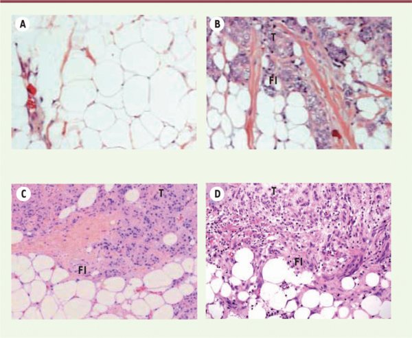
Figure 4.
Télécharger l'image originale
La proximité entre tissu adipeux et cellules tumorales est observée dans de nombreux cancers invasifs. Coupe histologique d’un tissu adipeux mammaire normal ( A ), d’un carcinome mammaire ( B ), prostatique ( C ) et d’un mélanome ( D ) après coloration à l’hématoxyline-éosine. Les adipocytes localisés au front invasif (FI) de la tumeur (T) ont une taille et un contenu lipidique diminués par rapport à ceux du tissu adipeux normal.
Current usage metrics show cumulative count of Article Views (full-text article views including HTML views, PDF and ePub downloads, according to the available data) and Abstracts Views on Vision4Press platform.
Data correspond to usage on the plateform after 2015. The current usage metrics is available 48-96 hours after online publication and is updated daily on week days.
Initial download of the metrics may take a while.