")
")
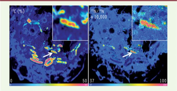
Figure 1.
Télécharger l'image originale
L’aspartate est accessible à M. tuberculosis dans les macrophages. Analyse par NanoSIMS de la colocalisation (flèche montrant une bactérie grossie dans l’insert) de M. tuberculosis marqué au 13C (gauche) dans un macrophage infecté, incubé en présence d’aspartate marqué au 15N (droite). L’échelle de couleur représente l’enrichissement en 13C (gauche) ou 15N (droite) (© Ting-Di Wu et Jean-Luc Guerquin-Kern, Institut Curie, laboratoire de microscopie ionique, Inserm U759, Orsay, France).
Current usage metrics show cumulative count of Article Views (full-text article views including HTML views, PDF and ePub downloads, according to the available data) and Abstracts Views on Vision4Press platform.
Data correspond to usage on the plateform after 2015. The current usage metrics is available 48-96 hours after online publication and is updated daily on week days.
Initial download of the metrics may take a while.