")
")
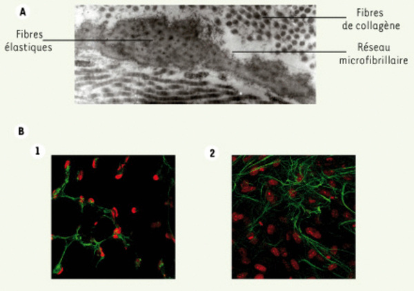
Figure 2.
Télécharger l'image originale
Le réseau fibrillaire. A. Visualisation par microscopie électronique du réseau microfibrillaire à partir d’une biopsie cutanée. Ce réseau forme une gangue autour des fibres élastiques pour former, ensemble, la matrice du tissu conjonctif élastique. B. Visualisation par immunofluorescence du réseau microfibrillaire au microscope confocal (x40).(1) Sans activation. (2) Après 5 jours d’exposition au TGF-β (10 ng/ml).
Current usage metrics show cumulative count of Article Views (full-text article views including HTML views, PDF and ePub downloads, according to the available data) and Abstracts Views on Vision4Press platform.
Data correspond to usage on the plateform after 2015. The current usage metrics is available 48-96 hours after online publication and is updated daily on week days.
Initial download of the metrics may take a while.