")
")
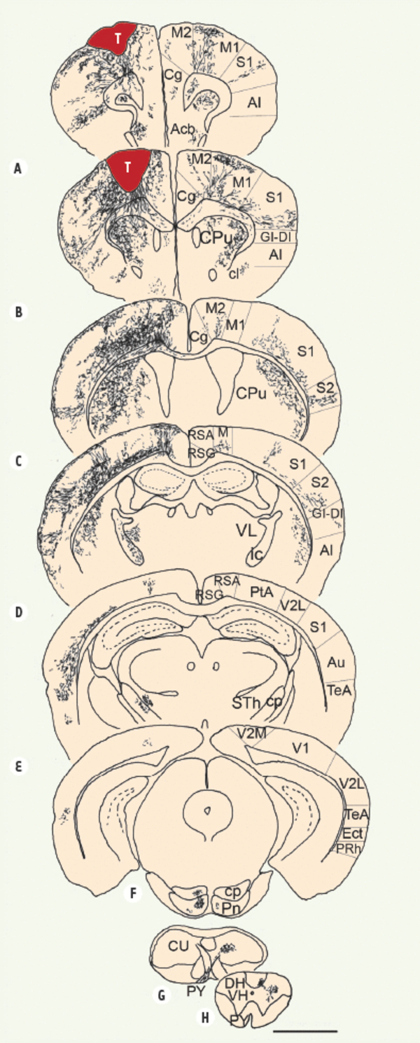
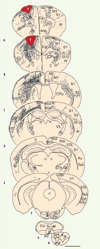
Figure 1.
Télécharger l'image originale
Représentation schématique de coupes frontales ordonnées selon l’axe rostrocaudal. Les panneaux A à H illustrent la distribution des fibres GFP+, 60 jours après greffe. Le transplant développe un ensemble de projections vers la plupart des cibles corticales et sous-corticales qui sont normalement contactées par les neurones du cortex moteur. Échelle : 600 µm. cc: corpus callosum ; cp : cerebral pedoncule ; CPu : caudate putamen ; dcs : dorsal corticospinal tract ; ic : internal capsule ; M : motor cortex ; Pn : pontine nuclei ; PY : pyramidal tract ; S : sensorimotor cortex ; Sth : subthalamic nucleus ; T : transplant ; VL : ventrolateral thalamic nucleus.
Current usage metrics show cumulative count of Article Views (full-text article views including HTML views, PDF and ePub downloads, according to the available data) and Abstracts Views on Vision4Press platform.
Data correspond to usage on the plateform after 2015. The current usage metrics is available 48-96 hours after online publication and is updated daily on week days.
Initial download of the metrics may take a while.