")
")
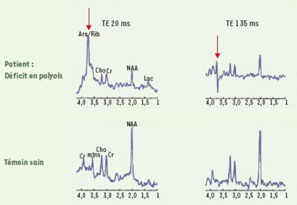
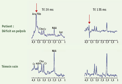
Figure 3.
Télécharger l'image originale
Spectroscopie IRM cérébrale dans les déficits héréditaires du métabolisme des polyols. Chez le patient, on note la présence d’un pic anormal (indiqué par la flèche) de D-arabitol et ribitol sur la séquence de spectroscopie à TE (temps d’écho) court (20 ms), correspondant à l’accumulation anormale de polyols cérébraux. Un pic anormal est également retrouvé sur la séquence à TE long (135 ms). Ara: D-arabitol; Rib: ribitol; Cho: choline; Cr: créatine; NAA: N-acétylaspartate; Lac: lactate; mIns: myoinositol (d’après [11]).
Current usage metrics show cumulative count of Article Views (full-text article views including HTML views, PDF and ePub downloads, according to the available data) and Abstracts Views on Vision4Press platform.
Data correspond to usage on the plateform after 2015. The current usage metrics is available 48-96 hours after online publication and is updated daily on week days.
Initial download of the metrics may take a while.