")
")
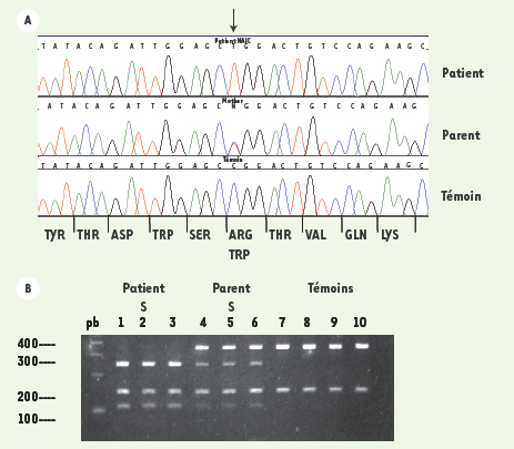
Figure 2.

Télécharger l'image originale
A. Profils d’électrophorèse des ADN d’un patient atteint de la cirrhose amérindienne, un porteur obligatoire et un individu témoin. Les acides aminés correspondant à la séquence de l’ADN apparaissent au bas de la figure. La mutation induit le remplacement d’une molécule d’arginine par une molécule de tryptophane.B. La détection de la mutation par PCR-RFLP [6]. La mutation crée un site de restriction pour l’enzyme de restriction Alu I. L’exon 15 du gène CIRH1A est amplifié par PCR. Après la digestion avec Alu I, les ADN sont séparés par électrophorèse sur un gel d’agarose 3 %. Le fragment amplifié par PCR contient un site de restriction constant Alu I. Celui-ci sert de contrôle positif de la digestion et donne un fragment de 150 pb. Puits 1-3 : échantillons des patients. Puits 4-6 échantillons des hétérozygotes et puits 7-10 échantillons des individus homozygotes normaux (pb : paires de base).
Current usage metrics show cumulative count of Article Views (full-text article views including HTML views, PDF and ePub downloads, according to the available data) and Abstracts Views on Vision4Press platform.
Data correspond to usage on the plateform after 2015. The current usage metrics is available 48-96 hours after online publication and is updated daily on week days.
Initial download of the metrics may take a while.